Correlative Atom probe tomography
Correlative atom probe tomography and electron microscopy refers to the combined utilization of a range of microscopy probing techniques to the same region in a single specimen.
This approach is increasingly deployed to understand fundamental aspects in nanoscale material science.
Particularly the combination of transmission electron microscopy (TEM) and atom probe tomography (APT) enables researchers to relate the atomic structure and composition of nanoscale features of interest and has been gaining influence over the past decades.
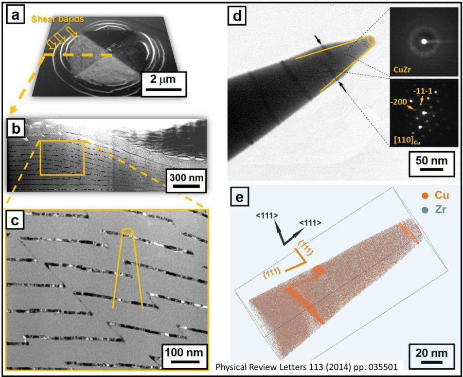 Correlative use of atom probe tomography (LEAP) in conjunction with electron microscopy for revealing both the structure and the chemical composition of a nanolaminate Cu Zr multilayer material (W. Guo, E. A. Jägle, P.-P. Choi, J. Yao, A. Kostka, J. M. Sc
PRL 113, 035501 (2014) PHYSICAL REVIEW LETTERS
Guo Raabe PHYSICAL REVIEW LETTERS vol 11[...]
PDF-Dokument [2.1 MB]
Acta Materialia 80 (2014) 94–106
Acta Materialia 80 (2014) 94 Nanolaminat[...]
PDF-Dokument [3.9 MB]
What is correlative electron microscopy and atom probe tomography ?
The correlative use of electron microscopy and atom probe tomography describe an experimental probing methodology which aims at revealing both, all relevant structural features and the chemical
composition at exactly the same material position in three dimensions at full atomic scale at ppm chemcial precision. This method is also sometimes referred to as correlative atom probe
tomography.
Typically the method works by preparing needle shaped tips with a tip apex radius of around 50 nm that are suited for atom probe tomography, yet, before doing so these tips are first exposed to
electron microscopical observations.
Then the two data sets from electron microscopy and atom probe tomography or jointly analysed.
Currently this combination of methods applied to exactly the same material portion represents the highest resolving joint crystallographic and chemical analysis method that can be applied to materials in 3D.
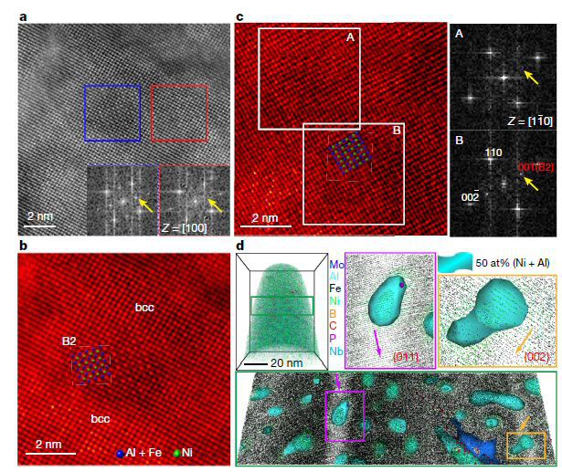 Coupled investigation of nanoparticles in a steel matrix conducted by using correlated electron microscopy and atom probe tomography, S. Jiang, H. Wang, Y. Wu, X. Liu, H. Chen, M. Yao, B. Gault, D. Ponge, D. Raabe, A. Hirata, M. Chen, Y. Wang, Z. Lu, Ultr
460 | NATURE | VOL 544 | 27 April 2017
Ultrastrong steel via minimal lattice mi[...]
PDF-Dokument [6.2 MB]
Why is correlative electron microscopy and atom probe tomography important ?
Using correlative electron microscopy and atom probe tomography enables us to reveal complex structural phenomena in materials and their interplay with chemical features. Most structural effects in complex modern materials, be at the formation of certain phases or the vast hierarchy of different types of lattice defects such as point defects, dislocations, grain boundaries or hetero-phase interfaces are characterized by specific chemical features. Therefore a more holistic understanding of nanostructured materials requires to reveal both, the local chemical composition together with the local structure of phases and defects to highest possible resolution in three dimensions.
 Atomic-Scale Quantification of Grain Boundary Segregation in Nanocrystalline Material; PRL 112, 126103 (2014) PHYSICAL REVIEW LETTERS 28 MARCH 2014
PRL 112, 126103 (2014) PHYSICAL REVIEW LETTERS 28 MARCH 2014
Phys Rev Lett. 2014 grain boundary segre[...]
PDF-Dokument [842.8 KB]
Ultramicroscopy vol 153 (2015) pages 32–39
APT Herbig Ultramicroscopy correlative m[...]
PDF-Dokument [3.3 MB]
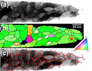
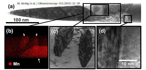 TEM analysis performed on APT tip. (a) BF-STEM micrograph of a martensitic Fe–9Mn alloy containing Mn-enriched precipitates. (b) STEM-EDX analysis of the Mn distribution (c) BF-TEM magnification (d) HRTEM image of austenite / martensite
Current Opinion in Solid State and Materials Science 18 (2014) 253–261
Current Opinion in Solid State Materials[...]
PDF-Dokument [1.7 MB]
 Combining structural and chemical information at the nanometer scale by correlative transmission electron microscopy and atom probe tomography on nanocrystalline iron.
Acta Materialia 84 (2015) 110–123
Acta Materialia 84 (2015) 110-123 atom p[...]
PDF-Dokument [2.4 MB]
Acta Materialia 59 (2011) 3965–3977
Acta Materialia 59 (2011) 3965 pearlite [...]
PDF-Dokument [1.0 MB]
What is the advantage of correlative electron microscopy and atom probe tomography compared to crystallographic atom probe tomography ?
Crystallographic atom probe refers to a method where some of the lattice planes can be resolved without the use of electron microscopy only through the adequate analysis of field desorption poles
of crystallographic nature from atom probe tomography directly.
However, different from electron microscopy in many atom probe tomographic experiments only some of the lattice planes can be resolved so that typically a full local crystallographic analysis is not
always possible.
Therefore a combined application of electron microscopy and atom probe tomography to the same sample region gives a more complete structural analysis then using crystallographic atom probe tomography
alone.
 In some cases crystallographic lattice planes can be directly resolved from atom probe tomography alone without the aid of electron microscopical methods.
Correlative Atom Probe Tomography and Electron Microscopy on Superalloys
Nanoscale solute segregation to or near lattice defects is a coupled diffusion
and trapping phenomenon that occurs in superalloys at high temperatures
during service. Understanding the mechanisms underpinning this crucial
process will open pathways to tuning the alloy composition for improving the
high-temperature performance and lifetime. In this project we introduce an approach combining atom probe tomography with high-end scanning electron microscopy techniques, in transmission
and backscattering modes, to enable direct investigation of solute segregation to defects generated during high-temperature deformation such as dislocations in a heat-treated Ni-based
superalloy and planar faults in a CoNi-based superalloy. In our project three different protocols were elaborated to capture the complete structural and compositional nature of the
targeted defect in the alloy.
 Correlative Atom Probe Tomography and Electon Microscopy (ECCI).
JOM 2018
https://doi.org/10.1007/s11837-018-2802-7
JOM 2018 Correlative Microscopy Makineni[...]
PDF-Dokument [2.4 MB]
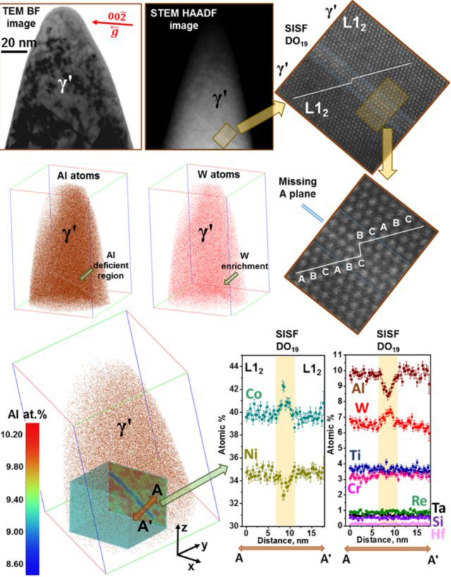 Correlative Microscopy: Novel Methods and Their Applications to Explore 3D Chemistry and Structure of Nanoscale Lattice Defects, here applied to superalloys (JOM, Vol. 70, No. 9, 2018).
Materialia 4 (2018) 109-114
Materialia 2018 Segregation Re dislocati[...]
PDF-Dokument [1.8 MB]
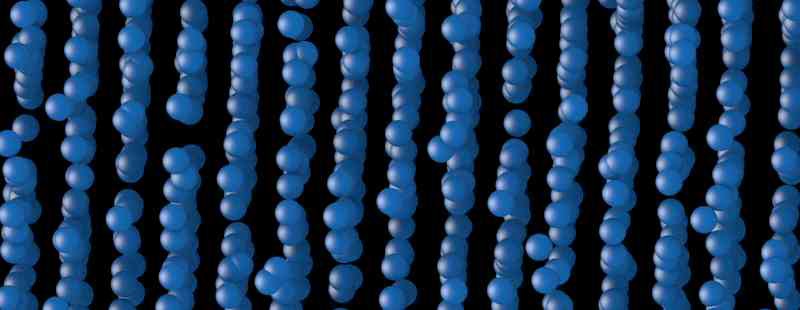
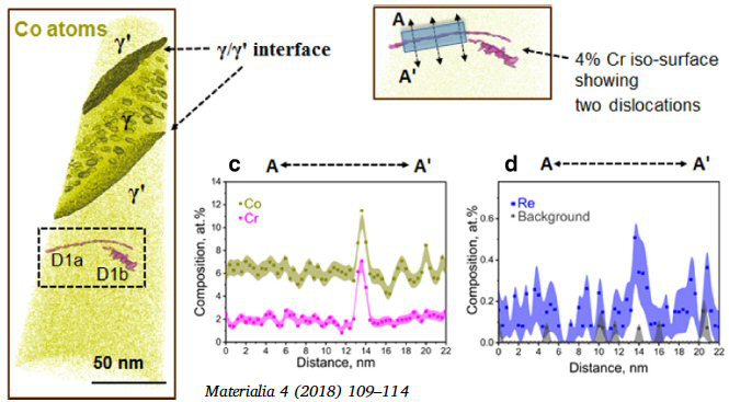 Ni-base superalloy: APT reconstruction of a tip prepared from the 1% sample showing a ?-channel between two ?/ ?’ interfaces. Two dislocations D1a and D1b are highlighted in the dashed black rectangle region.
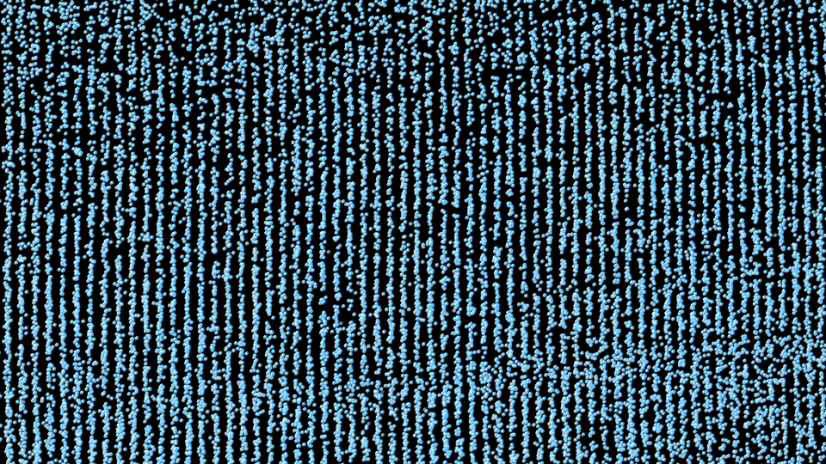
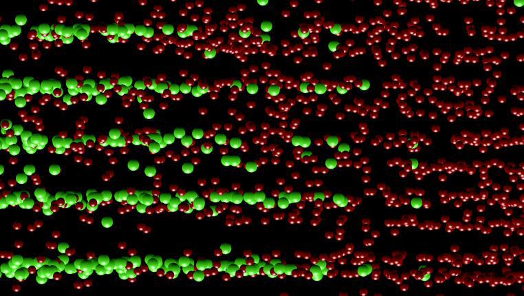
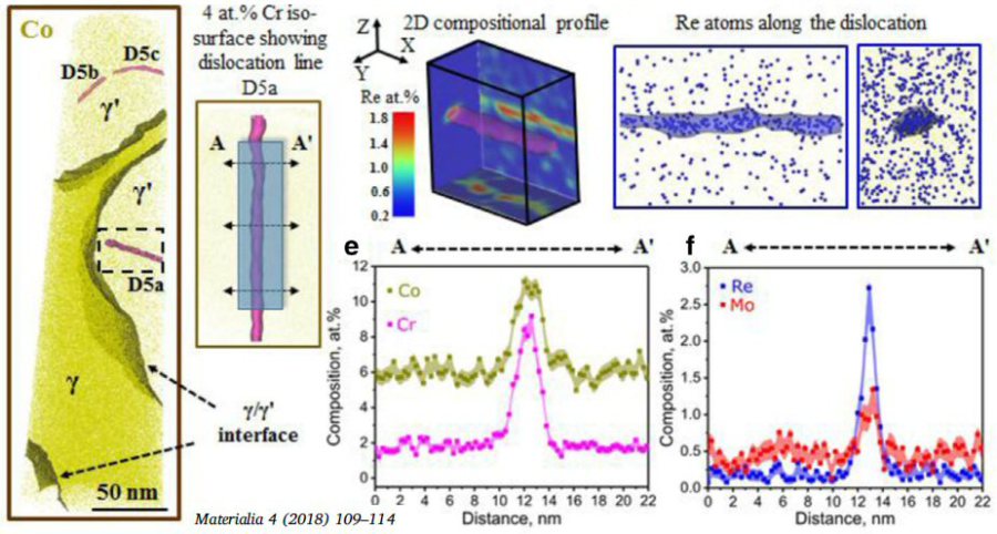 Rhenium in Ni-base superalloys: APT reconstruction of 5% deformation tip. Three dislocations are numbered as D5a, D5b and D5c. (b) Enlarged view of dislocation D5a. Composition profiles were measured in the marked region from A to A’.
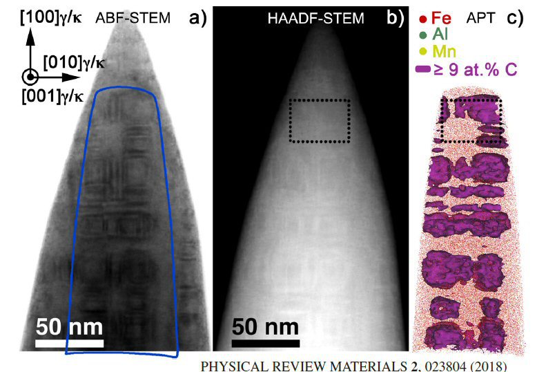
Atomistically resolved correlative scanning transmission electron microscopy and atom probe tomography
In this project correlative scanning transmission electron microscopy, atom probe tomography, and density functional theory calculations are jointly used to resolve the correlation between elastic
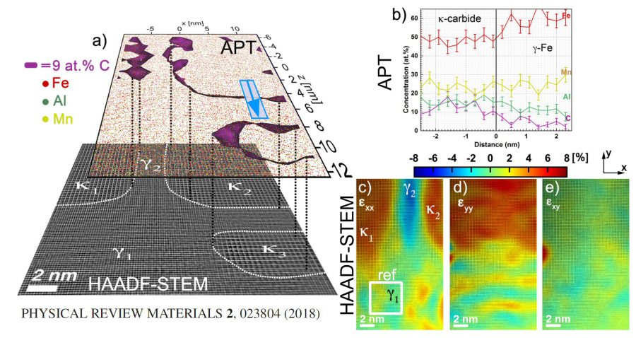
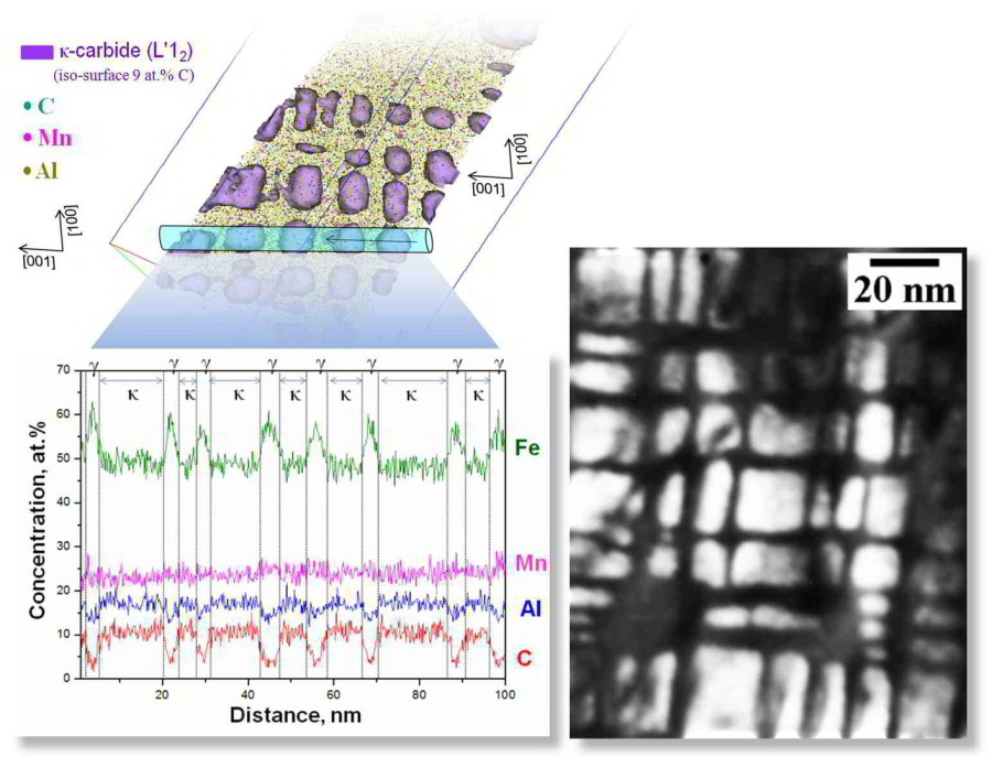
strain fields and local impurity concentrations on the atomic scale. The correlative approach is applied to coherent interfaces in a κ-carbide strengthened low-density steel and establishes
a tetragonal distortion of fcc-Fe. An interfacial roughness of ∼1 nm and a localized carbon
concentration gradient extending over ∼2–3 nm is revealed, which originates from the mechano-chemical coupling between local strain and composition.
PHYSICAL REVIEW MATERIALS 2, 023804 (2018)
PHYSICAL REVIEW MATERIALS 2 023804 (2018[...]
PDF-Dokument [1.4 MB]
 Atomistically resolved correlative scanning transmissin electron microscopy and atom probe tomography: PHYSICAL REVIEW MATERIALS 2, 023804 (2018)
 Atomistically resolved correlative scanning transmissin electron microscopy and atom probe tomography: PHYSICAL REVIEW MATERIALS 2, 023804 (2018)
Can we couple even high resolution (atomic column resolution) electron microscopy with atom probe tomography ?
Atom probe tomography can also be directly coupled with high resolution electron microscopy to resolve discrete atomic columns. The reason for this is that the needle shaped tips that are produced by using focused ion beam method for instance are so thin so that scanning transmission electron microscopy can resolve the atomic columns in such atom probe specimens prior to evaporation. Therefore atom probe tomography can be at the same region of interest directly combined with atomically resolving scanning transmission electron microscopy for resolving structural features in concert with chemical features at atomic scale.

 Combining high resolution scanning transmission electron microscopy with atom probe tomography, Acta Materialia 140 (2017) 258-273.