Field Ion Microscopy - Mapping single atoms
What is a Field Ion Microscope and where is it used?
The field ion microscope (FIM) is an atomically resolving analytical instrument used mainly in materials science.
It is a special microscope that makes the atomic arrangement on the surface of a sharp needle tip visible.
The method, which for the first time made atoms individually visible, was presented by Erwin Müller as a further development of his field electron microscope. Images of the atomic structure
of tungsten were first published in the Zeitschrift für Physik in 1951 (Erwin Wilhelm Müller: Das Feldionenmikroskop, Z. Phys. 131, 136 (1951)).
How does the Field Ion Microscope work?
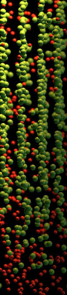
To examine a sample in a field ion microscope, a sharp metal tip is manufactured and placed in a vacuum chamber filled with a noble gas (e.g. helium or neon). Noble gases are used because a higher electric field is needed to ionize them due to their saturated and stable outer electron shells. Higher electric field then also means higher resolution. The tip is cooled down to a temperature between 20 and 100 K to reduce the random position changes of the atoms due to Brownian molecular motion. A positive high voltage of 5 to 10 kV is applied between the tip and a detector, e.g. a combination of microchannel plate and phosphor screen. In our group we also work directly with a multi-channel plate such as used in Atom Probe Tomography. A negative voltage at the tip would cause an undesired field emission of electrons.
Gas atoms are ionized by the strong electric field near the tip (therefore field ionization), positively charged and repelled by the tip. Although the electric field strength must be sufficiently
high to allow field ionization of the gas atoms, it must be low enough to avoid the detachment of atoms from the tip surface (field evaporation or field desorption).
In contrast to other microscope types (light microscope, electron microscope), where the magnification can be adjusted by means of optical elements (lenses), in the field ion microscope the
magnification depends essentially on the curvature of the tip surface and the applied voltage. The resolving power is not negatively influenced by aberrations of optical elements. There is
practically no wave-optical resolution limitation due to the low De-Broglie wavelength of the ions. The ionized gas atoms are accelerated radially (perpendicular to the surface) from the tip surface
onto the detector and provide a central projection of the tip surface with a magnification in the range of 100 Thousand to one Million. This makes it easy to image individual atoms of the tip surface
and to observe lattice defects such as dislocations.
What is the influence of the local electrostatic field rearrangement on field ionization in Field Ion Microscopy?
As described above field ion microscopy allows for direct imaging of surfaces with true atomic resolution. The high charge density distribution on the surface generates an intense
electric field that can induce ionization of gas atoms. In some projects we investigate the dynamic nature of the charge and the electrostatic field redistribution following the departure
of atoms initially
constituting the surface in the form of an ion, a process known as field evaporation. We developed a new algorithm for image processing and tracking of individual atoms on the specimen
surface enabling quantitative assessment of shifts in the imaged atomic positions. By combining experimental investigations with molecular dynamics simulations, which include the full
electric charge, we confirm that change is directly associated with the rearrangement of the electrostatic field that modifies the imaging gas ionization zone. We derive
important considerations for future developments of data reconstruction in 3D field ion microscopy, in particular for precise quantification of lattice strains and characterization of
crystalline defects at the atomic scale.
J. Phys. D: Appl. Phys. 51 (2018) 105601
Katnagallu_2018_J._Phys._D__Appl._Phys._[...]
PDF-Dokument [5.0 MB]
Can we map individual solute atoms at crystalline imperfections in metals with chemcial sensitivity in Field Ion Microscopy?
Directly imaging all atoms constituting a material and, maybe more importantly, crystalline defects that dictate materials’ properties, remains a formidable challenge. Here, we propose a new
approach to chemistry-sensitive field-ion microscopy (FIM)combining FIM with time-of-flight mass-spectrometry (tof-ms). Elemental identification and correlation to FIM images enabled by data mining
of
combined tof-ms delivers a truly analytical-FIM (A-FIM). Contrast variations due to different chemistries is also interpreted from density-functional theory (DFT). A-FIM has true
atomic resolution and we demonstrate how the technique can reveal the presence of individual solute atoms at specific positions in the microstructure. The performance of this new technique
is showcased in revealing individual Re atoms at crystalline defects formed in Ni–Re binary alloy during creep deformation. The atomistic details offered by A-FIM allowed us to directly
compare our results with simulations, and to tackle a long-standing question of how Re extends lifetime of Ni-based superalloys in service at high-temperature.
Shyam Katnagallu et al 2019 New J. Phys. 21 123020
Katnagallu_2019_New_J._Phys._21_123020 I[...]
PDF-Dokument [1.2 MB]